Analyze a Specific Site
This interface can be used to select probe binding hotspots that overlap with a given ligand.
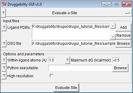
Input Files
Input files area list of ligand .pdb files and .dso file
from previous step. Ligand-bound form of the protein should be aligned
to the simulated structure. You can use prefix_heavyatoms.pdb
file for alignment.
Options & Parameters
- Parameters determined how many probe binding spots are incorporated in assessment of the specified site. Selected probe should be close to the ligand, so 1.5 Å should be sufficient. If the ligand is bound to a weak site, maximum free energy may be lower that that is used in the previous step, such as -0.5 kcal/mol.
- GUI will try to locate Python executable path, but if you do not see an entry, you will need to specify it manually.
When you use the tutorial files for MDM2 inhibitor, you should get a representation similar to the following:
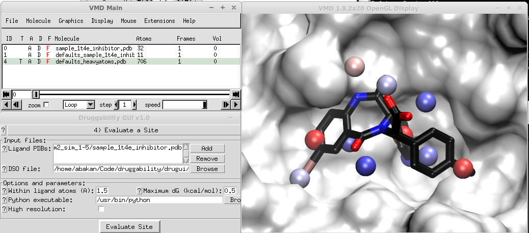
In the figure, 11 probe binding spots that overlap with MDM2 inhibitor is selected. Sum of their binding free energies looks reasonable (this will be displayed in the logfile viewer). For a large ligand, however, you may end up with a large selection of probe binding spots and sum of their binding free energies may result in a very high affinity. If this is the case, you should disregard the total. The approach (merging probe binding spots and adding their binding free energies) works well for drug-size molecules.
Output Files
Output from this step is a set of PDB files written into prefix
folder. Binding free energies of selected probe binding spots will
be appended to the log file in this folder.